Data Set Group2: GSE5281 Human Brain Normal Full Liang (Jul09)

|
|
| Contact Information |
Brandy Hamill
NIH Neuroscience Microarray Consortium
695 Charles E Young Dr S
Los Angeles, CA 90095 USA
Tel. 310-794-2416
bhamill@mednet.ucla.edu
Website
|
| Download datasets and supplementary data files |
|
|
|
|
| Specifics of this Data Set: |
None
|
| Summary: |
| (Taken verbatim from the GEO record)
Information about the genes that are preferentially expressed during the course of Alzheimer’s disease (AD) could improve our understanding of the molecular mechanisms involved in the pathogenesis of this common cause of cognitive impairment in older persons, provide new opportunities in the diagnosis, early detection, and tracking of this disorder, and provide novel targets for the discovery of interventions to treat and prevent this disorder. Information about the genes that are preferentially expressed in relationship to normal neurological aging could provide new information about the molecular mechanisms that are involved in normal age-related cognitive decline and a host of age-related neurological disorders, and they could provide novel targets for the discovery of interventions to mitigate some of these deleterious effects.
Aim 1. Collect brain samples from three Alzheimer’s Disease Centers (ADCs) for subsequent gene expression profiling. Individuals will be stratified with respect to diagnostic groups (using both clinical and neuropathological criteria), age groups, and APOE genotype. 150 individual brains will be sampled from the Arizona ADC, the Duke University ADC, and the Washington University ADC. Miniscule sample sizes (200 um of sectioned tissue) from six brain regions that are histopathologically or metabolically relevant to AD and aging will be collected, ensuring that this proposal does not deplete the national resource. Frozen and fixed samples will be sent to Phoenix, sectioned in a standardized fashion, and then returned. Aim 2. Tissue heterogeneity will be eliminated prior to expression profiling by performing laser capture microscopy on all brain regions. Aim 3. Expression profile LCM-captured cells on the Affymetrix U133 Plus 2.0 array (~55,000 transcripts), and quickly provide these data to the community at large. Aim 4. Identify pathogenic cascades related to each of the clinico-pathologic correlates using unsupervised and supervised analyses coupled with a hypothesis-driven approach. Aim 5. Validation of the expression correlates at the protein and functional levels.
Scientific progress in the last few years has improved our understanding of AD and raised the hope of identifying treatments to halt the progression and prevent the onset of this disorder. For instance, researchers have begun to characterize the cascade of molecular events which lead to the major histopathological features of the disorder: neuritic plaques, which contain extra-cellular deposits of amyloid beta-peptides (Abeta); neurofibrillary tangles, which contain the hyperphosphorylated form of the intracellular, microtubule-associated protein, tau; and a loss of neurons and synapses. These molecular events provide targets for the development of promising new treatments. For example, A-beta has been postulated to trigger a cascade of events that are involved in the pathogenesis of AD. This proposal hopes to provide new information about the genes that are preferentially expressed in the development of AD histopathology, including the over-expression of APP, amyloid-induced neurotoxicity, and hyperphosphorylation of tau, as well as bring clarity to the metabolic abnormalities that seem to play a role in dementia and AD development and pathology.
We will perform LCM on 6 brain regions with about 14 biological replicates per brain region. The brain regions are as follows: 1) entorhinal cortex 2) hippocampus 3) medial temporal gyrus 4) posterior cingulate 5) superior frontal gyrus and 6) primary visual cortex. We will collect layer III pyramidal cells from the white matter in each region, isolate total RNA from LCMed cell lysates, and perform double round amplification of each sample for array analysis.
Bad arrays excluded: Four samples, highlighted in the table below, are bad arrays. For quality control, they should be excluded.
| Index |
GEO Series |
Organ Region |
Tissue |
Case ID |
Age |
Sex |
| 1 |
GSM119615 |
Entorhinal Cortex |
Normal |
E119615M63N |
63 |
M |
| 2 |
GSM119616 |
Entorhinal Cortex |
Normal |
E119616M85N |
85 |
M |
| 3 |
GSM119617 |
Entorhinal Cortex |
Normal |
E119617M80N |
80 |
M |
| 4 |
GSM119618 |
Entorhinal Cortex |
Normal |
E119618M->F80N |
80 |
M |
| 5 |
GSM119619 |
Entorhinal Cortex |
Normal |
E119619F->M102N |
102 |
F |
| 6 |
GSM119620 |
Entorhinal Cortex |
Normal |
E119620M79N |
79 |
M |
| 7 |
GSM119621 |
Entorhinal Cortex |
Normal |
E119621M76N |
76 |
M |
| 8 |
GSM119622 |
Entorhinal Cortex |
Normal |
E119622M83N |
83 |
M |
| 9 |
GSM119623 |
Entorhinal Cortex |
Normal |
E119623M79N |
79 |
M |
| 10 |
GSM119624 |
Entorhinal Cortex |
Normal |
E119624F88N |
88 |
F |
| 11 |
GSM119625 |
Entorhinal Cortex |
Normal |
E119625F82N |
82 |
F |
| 12 |
GSM119626 |
Entorhinal Cortex |
Normal |
E119626M69N |
69 |
M |
| 13 |
GSM119627 |
Entorhinal Cortex |
Normal |
E119627M78N |
78 |
M |
| 14 |
GSM238763 |
Entorhinal Cortex |
Alzheimer's |
E238763F82A |
82 |
F |
| 15 |
GSM238790 |
Entorhinal Cortex |
Alzheimer's |
E238790F86A |
86 |
F |
| 16 |
GSM238791 |
Entorhinal Cortex |
Alzheimer's |
E238791F93A |
93 |
F |
| 17 |
GSM238792 |
Entorhinal Cortex |
Alzheimer's |
E238792M84A |
84 |
M |
| 18 |
GSM238793 |
Entorhinal Cortex |
Alzheimer's |
E238793F79A |
79 |
F |
| 19 |
GSM238794 |
Entorhinal Cortex |
Alzheimer's |
E238794F78A |
78 |
F |
| 20 |
GSM238795 |
Entorhinal Cortex |
Alzheimer's |
E238795F91A |
91 |
F |
| 21 |
GSM238796 |
Entorhinal Cortex |
Alzheimer's |
E238796M86A |
86 |
M |
| 22 |
GSM238797 |
Entorhinal Cortex |
Alzheimer's |
E238797NA0A |
N/A |
N/A |
| 23 |
GSM238798 |
Entorhinal Cortex |
Alzheimer's |
E238798M80A |
80 |
M |
| 24 |
GSM119628 |
Hippocampus |
Normal |
H119628M85N |
85 |
M |
| 25 |
GSM119629 |
Hippocampus |
Normal |
H119629M80N |
80 |
M |
| 26 |
GSM119630 |
Hippocampus |
Normal |
H119630M80N |
80 |
M |
| 27 |
GSM119631 |
Hippocampus |
Normal |
H119631F102N |
102 |
F |
| 28 |
GSM119632 |
Hippocampus |
Normal |
H119632M63N |
63 |
M |
| 29 |
GSM119633 |
Hippocampus |
Normal |
H119633M79N |
79 |
M |
| 30 |
GSM119634 |
Hippocampus |
Normal |
H119634M76N |
76 |
M |
| 31 |
GSM119635 |
Hippocampus |
Normal |
H119635M83N |
83 |
M |
| 32 |
GSM119636 |
Hippocampus |
Normal |
H119636M79N |
79 |
M |
| 33 |
GSM119637 |
Hippocampus |
Normal |
H119637F88N |
88 |
F |
| 34 |
GSM119638 |
Hippocampus |
Normal |
H119638F73N |
73 |
F |
| 35 |
GSM119639 |
Hippocampus |
Normal |
H119639M69N |
69 |
M |
| 36 |
GSM119640 |
Hippocampus |
Normal |
H119640M78N |
78 |
M |
| 37 |
GSM238799 |
Hippocampus |
Alzheimer's |
H238799F73A |
73 |
F |
| 38 |
GSM238800 |
Hippocampus |
Alzheimer's |
H238800M81A |
81 |
M |
| 39 |
GSM238801 |
Hippocampus |
Alzheimer's |
H238801M78A |
78 |
M |
| 40 |
GSM238802 |
Hippocampus |
Alzheimer's |
H238802M75A |
75 |
M |
| 41 |
GSM238803 |
Hippocampus |
Alzheimer's |
H238803F70A |
70 |
F |
| 42 |
GSM238804 |
Hippocampus |
Alzheimer's |
H238804F85A |
85 |
F |
| 43 |
GSM238805 |
Hippocampus |
Alzheimer's |
H238805F77A |
77 |
F |
| 44 |
GSM238806 |
Hippocampus |
Alzheimer's |
H238806M79A |
79 |
M |
| 45 |
GSM238807 |
Hippocampus |
Alzheimer's |
H238807M88A |
88 |
M |
| 46 |
GSM238808 |
Hippocampus |
Alzheimer's |
H238808M72A |
72 |
M |
| 47 |
GSM119641 |
Medial Temporal Gyrus |
Normal |
MT119641M85N |
85 |
M |
| 48 |
GSM119642 |
Medial Temporal Gyrus |
Normal |
MT119642M80N |
80 |
M |
| 49 |
GSM119643 |
Medial Temporal Gyrus |
Normal |
MT119643F102N |
102 |
F |
| 50 |
GSM119644 |
Medial Temporal Gyrus |
Normal |
MT119644M63N |
63 |
M |
| 51 |
GSM119645 |
Medial Temporal Gyrus |
Normal |
MT119645M79N |
79 |
M |
| 52 |
GSM119646 |
Medial Temporal Gyrus |
Normal |
MT119646M83N |
83 |
M |
| 53 |
GSM119647 |
Medial Temporal Gyrus |
Normal |
MT119647M79N |
79 |
M |
| 54 |
GSM119648 |
Medial Temporal Gyrus |
Normal |
MT119648F88N |
88 |
F |
| 55 |
GSM119649 |
Medial Temporal Gyrus |
Normal |
MT119649F82N |
82 |
F |
| 56 |
GSM119650 |
Medial Temporal Gyrus |
Normal |
MT119650F73N |
73 |
F |
| 57 |
GSM119651 |
Medial Temporal Gyrus |
Normal |
MT119651M69N |
69 |
M |
| 58 |
GSM119652 |
Medial Temporal Gyrus |
Normal |
MT119652M->F78N |
78 |
M |
| 59 |
GSM238809 |
Medial Temporal Gyrus |
Alzheimer's |
MT238809M81A |
81 |
M |
| 60 |
GSM238810 |
Medial Temporal Gyrus |
Alzheimer's |
MT238810M72A |
72 |
M |
| 61 |
GSM238811 |
Medial Temporal Gyrus |
Alzheimer's |
MT238811M75A |
75 |
M |
| 62 |
GSM238812 |
Medial Temporal Gyrus |
Alzheimer's |
MT238812M78A |
78 |
M |
| 63 |
GSM238813 |
Medial Temporal Gyrus |
Alzheimer's |
MT238813M75A |
75 |
M |
| 64 |
GSM238815 |
Medial Temporal Gyrus |
Alzheimer's |
MT238815F95A |
95 |
F |
| 65 |
GSM238816 |
Medial Temporal Gyrus |
Alzheimer's |
MT238816F81A |
81 |
F |
| 66 |
GSM238817 |
Medial Temporal Gyrus |
Alzheimer's |
MT238817F85A |
85 |
F |
| 67 |
GSM238818 |
Medial Temporal Gyrus |
Alzheimer's |
MT238818M79A |
79 |
M |
| 68 |
GSM238819 |
Medial Temporal Gyrus |
Alzheimer's |
MT238819F82A |
82 |
F |
| 69 |
GSM238820 |
Medial Temporal Gyrus |
Alzheimer's |
MT238820M88A |
88 |
M |
| 70 |
GSM238821 |
Medial Temporal Gyrus |
Alzheimer's |
MT238821M72A |
72 |
M |
| 71 |
GSM238822 |
Medial Temporal Gyrus |
Alzheimer's |
MT238822F73A |
73 |
F |
| 72 |
GSM238823 |
Medial Temporal Gyrus |
Alzheimer's |
MT238823M87A |
87 |
M |
| 73 |
GSM238824 |
Medial Temporal Gyrus |
Alzheimer's |
MT238824M68A |
68 |
M |
| 74 |
GSM238825 |
Medial Temporal Gyrus |
Alzheimer's |
MT238825F80A |
80 |
F |
| 75 |
GSM119653 |
Posterior Cingulate |
Normal |
PC119653M85N |
85 |
M |
| 76 |
GSM119654 |
Posterior Cingulate |
Normal |
PC119654M80N |
80 |
M |
| 77 |
GSM119655 |
Posterior Cingulate |
Normal |
PC119655F102N |
102 |
F |
| 78 |
GSM119656 |
Posterior Cingulate |
Normal |
PC119656M63N |
63 |
M |
| 79 |
GSM119657 |
Posterior Cingulate |
Normal |
PC119657M79N |
79 |
M |
| 80 |
GSM119658 |
Posterior Cingulate |
Normal |
PC119658M->F76N |
76 |
M |
| 81 |
GSM119659 |
Posterior Cingulate |
Normal |
PC119659M83N |
83 |
M |
| 82 |
GSM119660 |
Posterior Cingulate |
Normal |
PC119660M79N |
79 |
M |
| 83 |
GSM119661 |
Posterior Cingulate |
Normal |
PC119661F88N |
88 |
F |
| 84 |
GSM119662 |
Posterior Cingulate |
Normal |
PC119662F82N |
82 |
F |
| 85 |
GSM119663 |
Posterior Cingulate |
Normal |
PC119663F73N |
73 |
F |
| 86 |
GSM119664 |
Posterior Cingulate |
Normal |
PC119664M69N |
69 |
M |
| 87 |
GSM119665 |
Posterior Cingulate |
Normal |
PC119665M78N |
78 |
M |
| 88 |
GSM238826 |
Posterior Cingulate |
Alzheimer's |
PC238826F73A |
73 |
F |
| 89 |
GSM238827 |
Posterior Cingulate |
Alzheimer's |
PC238827M81A |
81 |
M |
| 90 |
GSM238834 |
Posterior Cingulate |
Alzheimer's |
PC238834M78A |
78 |
M |
| 91 |
GSM238835 |
Posterior Cingulate |
Alzheimer's |
PC238835M75A |
75 |
M |
| 92 |
GSM238837 |
Posterior Cingulate |
Alzheimer's |
PC238837M68A |
68 |
M |
| 93 |
GSM238838 |
Posterior Cingulate |
Alzheimer's |
PC238838F70A |
70 |
F |
| 94 |
GSM238839 |
Posterior Cingulate |
Alzheimer's |
PC238839F85A |
85 |
F |
| 95 |
GSM238840 |
Posterior Cingulate |
Alzheimer's |
PC238840M79A |
79 |
M |
| 96 |
GSM238841 |
Posterior Cingulate |
Alzheimer's |
PC238841M88A |
88 |
M |
| 97 |
GSM119666 |
Superior Frontal Gyrus |
Normal |
SF119666M79N |
79 |
M |
| 98 |
GSM119667 |
Superior Frontal Gyrus |
Normal |
SF119667F->M88N |
88 |
F |
| 99 |
GSM119668 |
Superior Frontal Gyrus |
Normal |
SF119668F->M82N |
82 |
F |
| 100 |
GSM119669 |
Superior Frontal Gyrus |
Normal |
SF119669F->M73N |
73 |
F |
| 101 |
GSM119670 |
Superior Frontal Gyrus |
Normal |
SF119670F->M102N |
102 |
F |
| 102 |
GSM119671 |
Superior Frontal Gyrus |
Normal |
SF119671M63N |
63 |
M |
| 103 |
GSM119672 |
Superior Frontal Gyrus |
Normal |
SF119672M->F79N |
79 |
M |
| 104 |
GSM119673 |
Superior Frontal Gyrus |
Normal |
SF119673M->F76N |
76 |
M |
| 105 |
GSM119674 |
Superior Frontal Gyrus |
Normal |
SF119674M->F83N |
83 |
M |
| 106 |
GSM119675 |
Superior Frontal Gyrus |
Normal |
SF119675M69N |
69 |
M |
| 107 |
GSM119676 |
Superior Frontal Gyrus |
Normal |
SF119676M78N |
78 |
M |
| 108 |
GSM238842 |
Superior Frontal Gyrus |
Alzheimer's |
SF238842F73A |
73 |
F |
| 109 |
GSM238843 |
Superior Frontal Gyrus |
Alzheimer's |
SF238843M81A |
81 |
M |
| 110 |
GSM238844 |
Superior Frontal Gyrus |
Alzheimer's |
SF238844M72A |
72 |
M |
| 111 |
GSM238845 |
Superior Frontal Gyrus |
Alzheimer's |
SF238845M75A |
75 |
M |
| 112 |
GSM238846 |
Superior Frontal Gyrus |
Alzheimer's |
SF238846M78A |
78 |
M |
| 113 |
GSM238847 |
Superior Frontal Gyrus |
Alzheimer's |
SF238847M75A |
75 |
M |
| 114 |
GSM238848 |
Superior Frontal Gyrus |
Alzheimer's |
SF238848M87A |
87 |
M |
| 115 |
GSM238851 |
Superior Frontal Gyrus |
Alzheimer's |
SF238851F95A |
95 |
F |
| 116 |
GSM238854 |
Superior Frontal Gyrus |
Alzheimer's |
SF238854M68A |
68 |
M |
| 117 |
GSM238855 |
Superior Frontal Gyrus |
Alzheimer's |
SF238855F95A |
95 |
F |
| 118 |
GSM238856 |
Superior Frontal Gyrus |
Alzheimer's |
SF238856F70A |
70 |
F |
| 119 |
GSM238857 |
Superior Frontal Gyrus |
Alzheimer's |
SF238857F85A |
85 |
F |
| 120 |
GSM238858 |
Superior Frontal Gyrus |
Alzheimer's |
SF238858F83A |
83 |
F |
| 121 |
GSM238860 |
Superior Frontal Gyrus |
Alzheimer's |
SF238860F77A |
77 |
F |
| 122 |
GSM238861 |
Superior Frontal Gyrus |
Alzheimer's |
SF238861F83A |
83 |
F |
| 123 |
GSM238862 |
Superior Frontal Gyrus |
Alzheimer's |
SF238862M68A |
68 |
M |
| 124 |
GSM238863 |
Superior Frontal Gyrus |
Alzheimer's |
SF238863M79A |
79 |
M |
| 125 |
GSM238864 |
Superior Frontal Gyrus |
Alzheimer's |
SF238864F82A |
82 |
F |
| 126 |
GSM238865 |
Superior Frontal Gyrus |
Alzheimer's |
SF238865M88A |
88 |
M |
| 127 |
GSM238867 |
Superior Frontal Gyrus |
Alzheimer's |
SF238867F80A |
80 |
F |
| 128 |
GSM238868 |
Superior Frontal Gyrus |
Alzheimer's |
SF238868M74A |
74 |
M |
| 129 |
GSM238870 |
Superior Frontal Gyrus |
Alzheimer's |
SF238870M72A |
72 |
M |
| 130 |
GSM238871 |
Superior Frontal Gyrus |
Alzheimer's |
SF238871M80A |
80 |
M |
| 131 |
GSM119677 |
Primary Visual Cortex |
Normal |
V119677M85N |
85 |
M |
| 132 |
GSM119678 |
Primary Visual Cortex |
Normal |
V119678M80N |
80 |
M |
| 133 |
GSM119679 |
Primary Visual Cortex |
Normal |
V119679M63N |
63 |
M |
| 134 |
GSM119680 |
Primary Visual Cortex |
Normal |
V119680M79N |
79 |
M |
| 135 |
GSM119681 |
Primary Visual Cortex |
Normal |
V119681M76N |
76 |
M |
| 136 |
GSM119682 |
Primary Visual Cortex |
Normal |
V119682M83N |
83 |
M |
| 137 |
GSM119683 |
Primary Visual Cortex |
Normal |
V119683M79N |
79 |
M |
| 138 |
GSM119684 |
Primary Visual Cortex |
Normal |
V119684F88N |
88 |
F |
| 139 |
GSM119685 |
Primary Visual Cortex |
Normal |
V119685F82N |
82 |
F |
| 140 |
GSM119686 |
Primary Visual Cortex |
Normal |
V119686F73N |
73 |
F |
| 141 |
GSM119687 |
Primary Visual Cortex |
Normal |
V119687M69N |
69 |
M |
| 142 |
GSM119688 |
Primary Visual Cortex |
Normal |
V119688M78N |
78 |
M |
| 143 |
GSM238872 |
Primary Visual Cortex |
Alzheimer's |
V238872F73A |
73 |
F |
| 144 |
GSM238873 |
Primary Visual Cortex |
Alzheimer's |
V238873M81A |
81 |
M |
| 145 |
GSM238874 |
Primary Visual Cortex |
Alzheimer's |
V238874M75A |
75 |
M |
| 146 |
GSM238875 |
Primary Visual Cortex |
Alzheimer's |
V238875M78A |
78 |
M |
| 147 |
GSM238877 |
Primary Visual Cortex |
Alzheimer's |
V238877M75A |
75 |
M |
| 148 |
GSM238941 |
Primary Visual Cortex |
Alzheimer's |
V238941M87A |
87 |
M |
| 149 |
GSM238942 |
Primary Visual Cortex |
Alzheimer's |
V238942F95A |
95 |
F |
| 150 |
GSM238943 |
Primary Visual Cortex |
Alzheimer's |
V238943M68A |
68 |
M |
| 151 |
GSM238944 |
Primary Visual Cortex |
Alzheimer's |
V238944F70A |
70 |
F |
| 152 |
GSM238945 |
Primary Visual Cortex |
Alzheimer's |
V238945F81A |
81 |
F |
| 153 |
GSM238946 |
Primary Visual Cortex |
Alzheimer's |
V238946F85A |
85 |
F |
| 154 |
GSM238947 |
Primary Visual Cortex |
Alzheimer's |
V238947M68A |
68 |
M |
| 155 |
GSM238948 |
Primary Visual Cortex |
Alzheimer's |
V238948M79A |
79 |
M |
| 156 |
GSM238949 |
Primary Visual Cortex |
Alzheimer's |
V238949F82A |
82 |
F |
| 157 |
GSM238951 |
Primary Visual Cortex |
Alzheimer's |
V238951M88A |
88 |
M |
| 158 |
GSM238952 |
Primary Visual Cortex |
Alzheimer's |
V238952M74A |
74 |
M |
| 159 |
GSM238953 |
Primary Visual Cortex |
Alzheimer's |
V238953M72A |
72 |
M |
| 160 |
GSM238955 |
Primary Visual Cortex |
Alzheimer's |
V238955M->F68A |
68 |
M |
| 161 |
GSM238963 |
Primary Visual Cortex |
Alzheimer's |
V238963F80A |
80 |
F |
|
|
| About the cases used to generate this set of data: |
|
| About the tissue used to generate this set of data: |
|
| About the array platform: |
| Affymetrix submissions are typically submitted to GEO using the GEOarchive method described at http://www.ncbi.nlm.nih.gov/projects/geo/info/geo_affy.html Complete coverage of the Human Genome U133 Set plus 6,500 additional genes for analysis of over 47,000 transcripts All probe sets represented on the GeneChip Human Genome U133 Set are identically replicated on the GeneChip Human Genome U133 Plus 2.0 Array. The sequences from which these probe sets were derived were selected from GenBank®, dbEST, and RefSeq. The sequence clusters were created from the UniGene database (Build 133, April 20, 2001) and then refined by analysis and comparison with a number of other publicly available databases, including the Washington University EST trace repository and the University of California, Santa Cruz Golden-Path human genome database (April 2001 release).
In addition, there are 9,921 new probe sets representing approximately 6,500 new genes. These gene sequences were selected from GenBank, dbEST, and RefSeq. Sequence clusters were created from the UniGene database (Build 159, January 25, 2003) and refined by analysis and comparison with a number of other publicly available databases, including the Washington University EST trace repository and the NCBI human genome assembly (Build 31).
|
| About data values and data processing: |
|
| Notes: |
|
| Experiment Type: |
| Human brain expression data in patients with Alzheimer's disease and age-matched elderly control subjects. This cortical expression data set is taken from GEO GSE5281 (Liang et al. 2006, Liang et al. 2008). Samples were laser-captured from cortical regions of 16 normal elderly humans (10 males and 4 females) and from 33 AD cases (15 males and 18 females). Mean age of cases and controls was 80 years. All samples were run on the Affymetrix U133 Plus 2.0 array. We renormalized the RMA data to an average expression of 8 units on a log2 scale. Two versions of the data have been entered in GeneNetwork: one consisting of 157 of 161 arrays (full set minus 4 arrays we consider of poor quality); the second consisting of what we regard as the best 102 arrays (those with mean correlations of better than 0.88 with all other arrays). Case IDs have the following code structure: Brain Region, GEO ID, Sex, Age, Disease Status. E119615M63N is a sample of the entorhinal cortex of case GSM119615, a male 63 year old normal case. The tissue codes are E = enorhinal cortex layer II, H = hippocampus CA1 pyramidal layer, MT = medial temporal cortex layer III, PC = porterior cingulate cortex layer III, SP = supeior frontal cortex layer III, V = primary visual cortex area 17 layer III. GeneNetwork does not yet allow sophisticated display of the data, but you can perform correlation analyses of any of the 56,000 probe sets. For example, expression of the APP transcript is higher in the AD cases and correlates well with many other AD related genes.
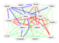
NOTE: We detected a minimum of 7.6% case assignment error rate (12 of 158 arrays) in this data set. Twelve cases are assigned to the wrong sex (see XIST probe set 224588_at, the figure below, and table 1). This raises the possibility that some cases are also misassigned by cortical brain region and disease status.

Legend: Expression of the sex-specific gene XIST reveals about 10 sex assignment errors in this data set.
Samples were laser-captured from cortical layer 3 (except the hippocampus) and run on the Affymetrix U133 Plus 2.0 array. We renormalized the data to an average expression of 8 units on a log2 scale. Case IDs have the following code structure: Brain Region, GEO ID, Sex, Age, Disease Status. E119615M63N is a sample of the entorhinal cortex of case GSM119615, a male 63 year old normal case. The tissue codes are E = enorhinal cortex layer II, H = hippocampus CA1 pyramidal layer, MT = medial temporal cortex layer III, PC = porterior cingulate cortex layer III, SP = supeior frontal cortex layer III, V = primary visual cortex layer III. A total of 16 normal subjects were used (10 M and 4 female). The AD samples. GeneNetwork does not allow sophisticated display of the data, but you can perform correlation analyses of any of the 56,000 probe sets. For example expression of the APP transcript is higher in the AD cases and correlates well with many other AD related genes.
Information about the genes that are preferentially expressed during the course of Alzheimer's disease (AD) could improve our understanding of the molecular mechanisms involved in the pathogenesis of this common cause of cognitive impairment in older persons, provide new opportunities in the diagnosis, early detection, and tracking of this disorder, and provide novel targets for the discovery of interventions to treat and prevent this disorder. Information about the genes that are preferentially expressed in relationship to normal neurological aging could provide new information about the molecular mechanisms that are involved in normal age-related cognitive decline and a host of age-related neurological disorders, and they could provide novel targets for the discovery of interventions to mitigate some of these deleterious effects.
|
| Contributor: |
| Stephan DA, Liang WS
|
| Citation: |
| Liang WS, Dunckley T, Beach TG, Grover A et al. Gene expression profiles in anatomically and functionally distinct regions of the normal aged human brain. Physiol Genomics 2007 Feb 12;28(3):311-22. PMID: 17077275
Liang WS, Reiman EM, Valla J, Dunckley T et al. Alzheimer's disease is associated with reduced expression of energy metabolism genes in posterior cingulate neurons. Proc Natl Acad Sci U S A 2008 Mar 18;105(11):4441-6. PMID: 18332434
|
| Data source acknowledgment: |
| Please cite: Liang WS, Reiman EM, Valla J, Dunckley T, Beach TG, Grover A, Niedzielko TL, Schneider LE, Mastroeni D, Caselli R, Kukull W, Morris JC, Hulette CM, Schmechel D, Rogers J, Stephan DA (2008) Alzheimer's disease is associated with reduced expression of energy metabolism genes in posterior cingulate neurons. Proc Natl Acad Sci USA 105:4441-4446.
|
| Study Id: |
77
|
|
|

|
Web services initiated January, 1994 as Portable Dictionary of the Mouse Genome; June 15, 2001 as WebQTL; and Jan 5, 2005 as GeneNetwork.
This site is currently operated by
Rob Williams,
Pjotr Prins,
Zachary Sloan,
Arthur Centeno. Design and code by Pjotr Prins, Zach Sloan, Arthur Centeno, Danny Arends, Christian Fischer, Sam Ockman, Lei Yan, Xiaodong Zhou, Christian Fernandez, Ning Liu, Rudi Alberts, Elissa Chesler, Sujoy Roy, Evan G. Williams, Alexander G. Williams, Kenneth Manly, Jintao Wang, and Robert W. Williams, colleagues.
|

|

|
GeneNetwork support from:
- The UT Center for Integrative and Translational Genomics
- NIGMS Systems Genetics and Precision Medicine project (R01 GM123489, 2017-2021)
- NIDA NIDA Core Center of Excellence in Transcriptomics, Systems Genetics,
and the Addictome (P30 DA044223, 2017-2022)
- NIA Translational Systems Genetics of Mitochondria, Metabolism, and Aging (R01AG043930, 2013-2018)
- NIAAA Integrative Neuroscience Initiative on Alcoholism (U01 AA016662, U01 AA013499, U24 AA013513, U01 AA014425, 2006-2017)
- NIDA, NIMH, and NIAAA (P20-DA 21131, 2001-2012)
- NCI MMHCC (U01CA105417), NCRR, BIRN, (U24 RR021760)
|
|
It took 0.047 second(s) for tux01.uthsc.edu to generate this page
|
|